Motivation
High-quality segmentation typically requires expert-drawn masks and hours of annotation per study. Weak supervision reduces this burden by using coarser, cheaper signals during training while still targeting pixel-level outputs at inference. The thesis evaluates a weakly supervised pipeline for prostate lesion segmentation that minimizes manual effort without discarding rigor in evaluation.
Dataset
For our work, we used the publicly available Prostate158 dataset, consisting of 158 prostate MRI studies (T2w, DWI, ADC sequences). We used 139 cases for training and 19 for testing. The 139‑case development set was split into 122 training and 17 validation cases. Ground‑truth lesion masks were withheld from training and used only for validation and final test evaluation.

Methodology Overview
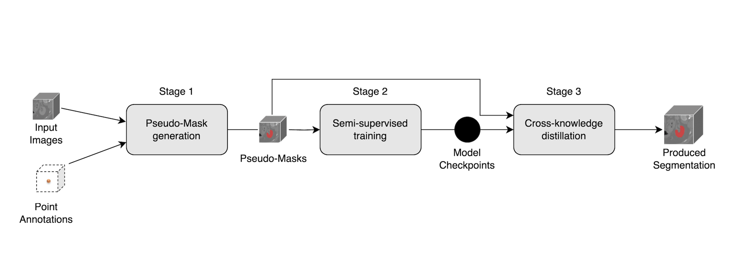
The pipeline comprises three stages designed to leverage minimal supervision while mitigating noise from automatically generated labels.
- Pseudo‑label generation via a pre‑trained model
- Single point prompts are automatically derived from reference masks (centroid-level guidance only).
- A foundation model adapted for volumetric data (SAM‑Med3D) converts these point prompts into volumetric pseudo‑segmentations.
- Semi‑supervised learning with complementary architectures
- Two segmentation models are trained on pseudo‑labels.
- 2.5D U‑Net (for anisotropic resolution and contextual slices)
- Attention U‑Net (attention gates to focus on salient features)
- Losses combine overlap- and distribution‑based terms to stabilize learning under pseudo‑label noise.
- Cross‑knowledge distillation
- After initial training, both models are fine‑tuned jointly using mutual supervision, where each model learns from the other’s predictions in addition to pseudo‑labels. This reduces overfitting to systematic errors in any single label source.
Evaluation
Evaluation follows standard segmentation metrics used in medical imaging.
- Dice similarity coefficient (primary)
- Hausdorff distance and average surface distance (boundary quality)

All metrics are reported on the held‑out test portion of Prostate158 using the original ground‑truth masks. No dense masks are used during training.
Results
- 2.5D U‑Net, Dice 0.404 on the Prostate158 test set
- Attention U‑Net, Dice 0.3097 on the Prostate158 test set
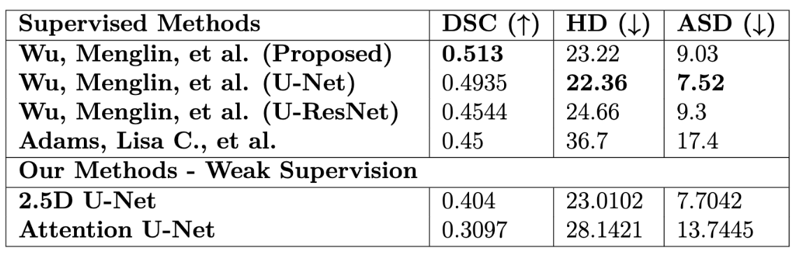
Qualitatively, the models delineate lesion regions with reasonable boundary adherence given weak supervision. Cross‑knowledge distillation improved robustness to pseudo‑label inaccuracies relative to training either model alone.
Discussion
- Annotation efficiency. Using a single point per study to drive pseudo‑label generation reduces labeling effort by orders of magnitude compared to dense masks.
- Model complementarity. Training two architectures and distilling between them counteracts bias from any one model or pseudo‑labeling artifact.
- Limitations. Performance remains below fully supervised approaches. Sensitivity to pseudo‑label quality persists. Further gains likely require improved prompting, uncertainty‑aware training, or stronger regularization of boundaries.
Conclusion
The study demonstrates that weak supervision, when paired with foundation‑model pseudo‑labels and cross‑model distillation, can yield competitive segmentation quality under stringent annotation budgets. This supports practical deployment paths where expert time is constrained, and provides a blueprint for extending the approach to other organs and modalities.
For technical details (data preprocessing, training schedules, and ablations), refer to the full thesis, Using weakly annotated data for medical image segmentation based on pre‑trained deep neural networks (May 2025).